At Synergy Health, we believe that the more our patients understand about the CCSVI procedure, the more comfortable they will be with the process. That’s why we provide extensive information about the procedure itself.
To help all of our patients understand what is done during the CCSVI procedure, we recorded an entire video of the procedure. Dr. Harris narrated the video to give patients an insight into how the procedure is performed. The following is a an actual CCSVI procedure:

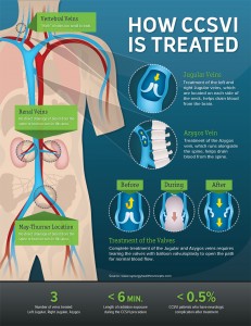
A comprehensive infographic showing how CCSVI is treated. We encourage you to copy and share this resource with others. Click to enlarge.
Treatment of central venous disease involves a variety of techniques. In the majority of cases, a large IV is placed into a larger vein in the groin area. From this location, small catheters are navigated into the neck and chest veins using fluoroscopy or X-ray guidance. The catheters are used to take pictures by injecting a dye into the veins to help identify any narrowing or blockage.
Sometimes these venograms do not adequately show the blockages. In these cases, our physicians use an ultrasound attached to the end of the small catheter to create pictures from inside of the veins. This technique increases the accuracy of the overall evaluation of the venous system and allows for more precise measurements of the veins themselves.
If a narrowing within the vein is seen, a small balloon can be placed across the narrowing and inflated. Angioplasty of the blockage can help open the narrowing and restore normal blood flow through the vein. Occasionally, balloon angioplasty does not improve the size of the vein and placement of a small stent is required. Placement of a stent can help open the vein back to its normal size.